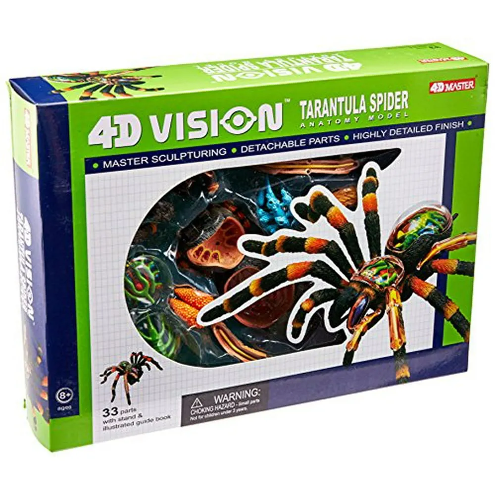
Understanding the 4D Tarantula Model
A 4D tarantula model offers an immersive and interactive way to explore the intricate anatomy of these fascinating creatures. Unlike static 2D diagrams, these models provide a three-dimensional view, allowing for a deeper understanding of how different body parts interact. The fourth dimension, often represented by interactivity or the ability to disassemble and reassemble parts, enhances the learning experience. These models are invaluable tools for students, educators, and anyone curious about spider anatomy. They provide a hands-on approach, making complex biological concepts easier to grasp and remember.
The Exoskeleton’s Role
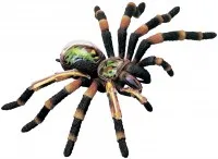
One of the most distinctive features of a tarantula is its exoskeleton a hard, protective outer shell. This exoskeleton serves several vital functions, including providing structural support, protection from predators, and preventing water loss. The exoskeleton is made of chitin, a tough, durable substance. The 4D model allows you to examine the exoskeleton in detail, seeing its various segments and the way it provides both flexibility and rigidity. The model also illustrates how the tarantula molts its exoskeleton as it grows, shedding the old shell to reveal a new, larger one.
Key Features of the Exoskeleton
The exoskeleton’s surface has numerous small hairs, or setae, which are important for sensory perception and provide the tarantula with information about its environment. The model can highlight the specific regions, like the carapace (the dorsal shield covering the cephalothorax) and the abdomen, providing an understanding of how these areas protect the vital organs. The model can also demonstrate the different sections and the way they are connected for movement.
Internal Anatomy Revealed
Beyond the exoskeleton lies a complex array of internal organs. A 4D model allows you to visualize these delicate structures in a way that 2D images simply cannot. It reveals the relative positions of organs and how they relate to one another within the body cavity. The model can be disassembled to provide a better understanding of the intricacies of the internal systems, making it an effective tool for learning about spider physiology.
The Digestive System
Tarantulas, like all spiders, have a unique digestive system. They inject enzymes into their prey to pre-digest it externally before sucking up the liquid nutrients. The model illustrates the key parts of the digestive tract, including the pharynx, the sucking stomach, and the intestines. You can follow the path of the food as it moves through the system, understanding how tarantulas efficiently extract nutrients from their meals. The model also highlights the filtering action within the digestive system, which screens out solid materials.
The Circulatory System
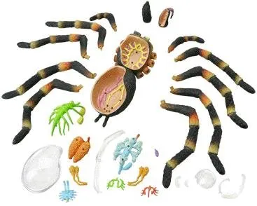
Tarantulas have an open circulatory system, where blood (hemolymph) flows freely within the body cavity, bathing the organs. The 4D model allows you to visualize the heart, a long, tubular organ located in the abdomen, and the vessels that carry hemolymph to the different parts of the body. Understanding the circulatory system helps you understand the transport of nutrients, oxygen, and waste products. The model can also reveal the ostia, small openings in the heart through which hemolymph enters.
The Nervous System
The tarantula’s nervous system is responsible for coordinating movement, sensing the environment, and controlling bodily functions. The 4D model shows the central nervous system, which consists of a brain (cerebral ganglia) and a ventral nerve cord that runs along the underside of the body. The model can highlight the ganglia that control specific functions, such as leg movement or sensory input. This provides a better understanding of how tarantulas react to their surroundings and make complex decisions.
Respiratory and Excretory Systems
The respiratory and excretory systems of a tarantula are key to its survival. The 4D model allows you to understand how these systems work to maintain internal balance. The model shows the crucial structures involved in breathing and waste removal.
Gas Exchange and Tracheae

Tarantulas breathe using book lungs, which are modified respiratory organs located in the abdomen. The 4D model shows the structure of the book lungs, including the lamellae, thin, plate-like structures that facilitate gas exchange. You can see how oxygen is absorbed from the air and how carbon dioxide is released. The model might also show the presence of tracheae, small tubes that carry air directly to the tissues.
Malpighian Tubules and Waste Removal
The tarantula’s excretory system relies on Malpighian tubules, which are slender tubes that filter waste products from the hemolymph. The 4D model shows how these tubules empty into the hindgut, where the waste is processed and expelled. This model helps users understand how the spider manages its waste products and maintains its internal environment.
Sensory Organs and Appendages
Tarantulas rely on a variety of sensory organs to navigate their environment, hunt prey, and avoid predators. A 4D model can highlight these sensory systems, allowing you to visualize how tarantulas interact with the world around them.
The Chelicerae and Pedipalps

The chelicerae are the tarantula’s mouthparts, used for grasping and injecting venom into prey. The 4D model can demonstrate the structure of the chelicerae, including the fangs, and their position. The pedipalps, located near the chelicerae, have sensory and manipulative functions. The model can demonstrate their role in sensing the environment.
Leg Structure and Function
Tarantula legs are incredibly complex structures. The 4D model demonstrates the segments of the legs including the coxa, trochanter, femur, patella, tibia, metatarsus, and tarsus. The model shows how these segments work together to provide the spider with movement. The legs have sensory hairs that allow the tarantula to detect vibrations and sense its environment.
Why 4D Models are Essential
4D tarantula models significantly enhance the learning experience by providing a dynamic and interactive understanding of spider anatomy. They allow for a more immersive and comprehensive exploration than static diagrams or text. They are invaluable tools for anyone interested in the anatomy of tarantulas, aiding in better comprehension and retention of complex biological concepts. The ability to visualize and manipulate a model of a tarantula’s anatomy fosters a deeper appreciation for these fascinating creatures and their survival strategies.